

Collection of 100+ important findings that are high yield for a medical student. This is important for USMLE Step 1 or other equivalent exams.
| 2 types of COPD |
Pink Puffer è Type A: Emphysema Blue Bloater è Type B: Bronchitis Emphysema- Centroacinar-smoking; Panacinar – α1-antitrypsin deficiency |
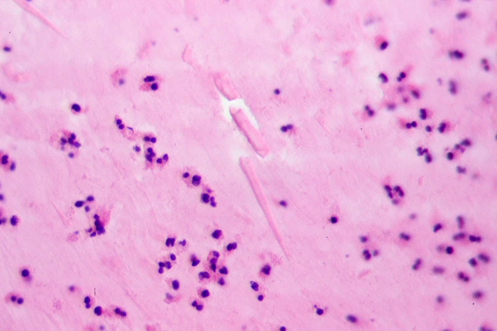
| 45 Degree Branch Points |
Aspergillosis |
| Acanthocytes |
RBSc w/ spiny projections. Seen in Abetalipoproteinemia. |
| Albumino-Cytologic Dissociation |
Guillain-Barre (markedly increased protein in CSF with only modest increase in cell count) |
| Antiplatelet Antibodies |
Idiopathic thrombocytopenic purpura |
| Arachnodactyly |
Marfan’s |
| Aschoff Bodies |
Rheumatic fever |
| Auer Rods |
Acute promyelocytic leukemia (AML type M3) |
| Autosplenectomy |
Sickle cell anemia: switch a glu ! val in β chain Low O2 ↑ sickling Aplastic crisis w/ B19 (Parvovirus ssDNA) infection Salmonella osteomyelitis Vaso-occlusive painful crisises Hydroxyurea as Txt (↑ HbF) & Bone marrow transplant |
| Babinski |
UMN lesion |
| Basophilic Stippling of RBCs |
Lead poisoning |
| Bence Jones Protein |
Multiple myeloma free light chains (either kappa or lambda) Waldenstrom’s macroglobinemia |
| Birbeck Granules |
Histiocytosis X (eosinophilic granuloma) |
| Blue Bloater |
Chronic Bronchitis (at least 3 months for at least 2 years of ecessive mucus secretion & chronic |
| Boot-Shaped Heart |
Tetralogy of Fallot |
| Both Sensory & Motor Lesion |
Brown Sequard; Anterior Spinal artery Occlusion |
| Both UMN & LMN Lesion |
ALS = Lou Gherig’s Disease |
| Bouchard’s Nodes |
Osteoarthritis (Proximal IP joint of the fingers) |
| Boutonniere’s Deformity |
Rheumatoid arthritis flex proximal & extend distal IP joints |
| Brown Tumor |
Hyperparathyroidism |
| Brushfield Spots |
Down’s |
| Call-Exner Bodies |
Granulosa cell tumor: associated w/ endometrial hyperplasia & carcinoma Granuloma-Theca cell tumor |
| Cardiomegaly with Apical Atrophy |
Chagas Disease |
| Chancre |
1° Syphilis |
| Chancroid |
Haemophilus ducreyi |
| Charcot Triad |
Multiple sclerosis = nystagmus, intention tremor, scanning speech |
| Charcot-Leyden Crystals |
Bronchial asthma |
| Cheyne-Stokes Breathing |
Cerebral lesion |
| Chocolate Cysts |
Endometriosis |
| Chvostek’s Sign |
Hypocalcemia facial spasm in tetany |
| Clue Cells |
Gardnerella vaginitis |
| Codman’s Triangle |
Osteosarcoma |
| Cold Agglutinins |
Mycoplasma pneumoniae Infectious mononucleosis |
| Condyloma Lata |
2° Syphilis |
| Congo Red |
Shows amyloid deposition in plaques & vascular walls |
| Cotton Wool Spots |
HTN; Aka, cytoid bodies seen w/ SLE (yellowish cotton wool fundal lesions) |
| Councilman Bodies |
Dying hepatocytes HepB |
| Cowdry A Inclusions |
Seen w/ Herpes Simplex Encephalitis in oligodendroglia |
| Crescents |
Goodpastures syndrome (pneumonia w/ hemoptysis & rapidly progressive glomerulonephritis) |
| Crescents In Bowman’s Capsule |
Rapidly progressive (crescentic glomerulonephritis) |
| Cuneocerebellar tr. |
Unconscious proprioception & fine motor movements of upper extremities |
| Currant-Jelly Sputum |
Klebsiella |
| Curschmann’s Spirals |
Bronchial asthma |
| Depigmentation Of Substantia Nigra |
Parkinson’s |
| Devic’s Syndrome |
Neuromyelitis Optica; A variant of multiple sclerosis: rapid demyelination of the optic nerve & spinal cord w/ paraplegia |
| Donovan Bodies |
Granuloma inguinale (STD) |
| Dorsal Column |
Conscious proprioception of the body |
| Dorsal Spinocerebellar tr. |
Unconscious prorpioception & fine motor movements |
| Eburnation |
Osteoarthritis (polished, ivory-like appearance of bone) |
| Ectopia Lentis |
Marfan’s |
| Erythema Chronicum Migrans |
Lyme Disease |
| Fatty Liver |
Alcoholism |
| Ferruginous Bodies |
Asbestosis – & Iron laden |
| Foster-Kennedy Syndrome |
A tumor causing blindness & loss of smell w/ papilloedema |
| Ghon Focus / Complex |
Tuberculosis (1° & 2°, respectively) |
| Glitter Cells |
Acute Pyelonephritis |
| Gower’s Maneuver |
Duchenne’s MD use of arms to stand |
| Ground Glass Appearance (Hyaline) |
Seen w/ Progressive Multifocal Leukoencephalopathy oligodendrocytes; Nuclei seen in Papillary CA of the thyroid (malignant) |
| Heberden’s Nodes |
Osteoarthritis (Distal IP joint of the fingers) |
| Heinz Bodies |
G6PDH Deficiency |
| Heterophil Antibodies |
Infectious mononucleosis (EBV) |
| Hirano Bodies |
Alzheimer’s |
| Hoffman’s Sign |
Flicking of the middle finger’s nail |
| Honey Combing of the lung |
Seen w/ Asbestosis (a restrictive lung disease) |
| Hypersegmented PMNs |
Megaloblastic anemia |
| Hypochromic Microcytic RBCs |
Iron-deficiency anemia or β Thalassemia |
| Jarisch-Herxheimer Reaction |
Syphilis over-aggressive treatment of an asymptomatic pt. that causes symptoms 2° to rapid lysis |
| Joint Mice |
Osteoarthritis (fractured osteophytes) |
| Kaussmaul Breathing |
Acidosis / Diabetic Ketoacidosis |
| Keratin Pearls |
Squamous Cell CA of skin Actinic Keratosis is a precursor |
| Keyser-Fleischer Ring |
Wilson’s |
| Kimmelstiel-Wilson Nodules |
Diabetic nephropathy: Nodular Glomerulosclerosis nodules of mesangial matrix |
| Koilocytes |
HPV 6 & 11 (condyloma acuminatum – benign) and HPV 16 & 18 (malignant association) |
| Koplik Spots |
Measles |
| LMN Lesion |
Werndig Hoffman (progressive infantile muscular atrophy); Poliomyelitis |
| Lateral Spinothalamic tr. |
Pain & Temperature sensation |
| Lewy Bodies |
Parkinson’s (eosinophilic inclusions in damaged substantia nigra cells) |
| Linear Ig Deposits |
Goodpastures syndrome |
| Lines of Zahn |
Arterial thrombus |
| Lisch Nodules |
Neurofibromatosis (von Recklinhausen’s disease) = pigmented iris hamartomas |
| Lumpy-Bumpy IF Glomeruli |
Poststreptococcal glomerulonephritis prototype of nephritic syndrome |
| Mallory Bodies |
Alcoholic hepatitis |
| Mamillary Body |
Can have hemorrhages as seen in Wernicke’s Encephalopathy |
| McBurney’s Sign |
Appendicitis (McBurney’s Point is 2/3 of the way from the umbilicus to anterior superior iliac spine) |
| Meningiomas & Progesterone |
Some meningiomas have Progesterone receptors = rapid growth in pregnancy can occur |
| Michealis-Gutmann Bodies |
Malakoplakia lesion on bladder due to macros & calcospherites (M-G Bodies): usually due to E. Coli |
| Monoclonal Antibody Spike |
Multiple myeloma this is called the M protein (usually IgG or IgA); MGUS |
| Myxedema |
Hypothyroidism |
| Negri Bodies |
Rabies |
| Neuritic Plaques |
Alzheimer’s |
| Neurofibrillary Tangles |
Alzheimer’s |
| Non-pitting Edema |
Myxedema; Anthrax Toxin |
| Notching of Ribs |
Coarctation of Aorta |
| Nutmeg Liver |
CHF = causing congested liver |
| Owls Eye Cells |
CMV; Reed Sternburg Cells (Hodkins Lymphoma); Aschoff cells seen w/ Rheumatic Fever |
| PAS(+) Dutcher Bodies |
Waldenstrom’s Macroglobulinemia = ↑IgM = Hyperviscosity |
| Painless Jaundice |
Pancreatic CA (head) |
| Pannus |
Rheumatoid arthritis, also see morning stiffnes that ↓ w/ joint use, HLA-DR4 |
| Pautrier’s Microabscesses |
Mycosis fungoides (cutaneous T-cell lymphoma), Sezary |
| Philadelphia Chromosome |
CML |
| Pick Bodies |
Pick’s Disease |
| Podagra |
Gout (MP joint of hallux) |
| Port-Wine Stain |
Hemangioma |
| Posterior Anterior Drawer Sign |
Tearing of the ACL |
| Psammoma Bodies |
Papillary adenocarcinoma of the thyroid Serous papillary cystadenocarcinoma of the ovary Meningioma Mesothelioma |
| Pseudohypertrophy |
Seen w/ Duchenne muscular dystrophy @ the claf muscles, due to ↑ fat |
| Punched-Out Bone Lesions |
Multiple myeloma |
| Rash on Palms & Soles |
2° Syphilis; RMSF; Coxsackie virus infection: Hand-Foot-Mouth Disease |
| Red Morning Urine |
Paroxysmal nocturnal hemoglobinuria. You would use Ham’s test to confirm. |
| Red Nucleus Destruction |
Intention tremors of the arm |
| Reed-Sternberg Cells |
Hodgkin’s Disease |
| Reid Index Increased |
Chronic bronchitis = ↑d ratio of bronchial gland to bronchial wall thickness |
| Reinke Crystals |
Leydig cell tumor |
| Rouleaux Formation |
Multiple myeloma RBC’s stacked as poker chips |
| S3 Heart Sound |
L→R Shunt (VSD, PDA, ASD); Mitral Regurg; LV Failure |
| S4 Heart Sound |
Pulmonary Stenosis; Pulmonary HTN |
| Schwartzman Reaction |
Neisseria meningitidis impressive rash with bugs |
| Sensory Pathway Lesion |
Subacute Combined Degeneration = Friedrich’s Ataxia = B12 deficiency; Tabes Dorsalis (Neurosyphilis |
| Smith Antigen |
SLE (also anti-dsDNA); Malar Rash, Wire loop kidney lesions, Joint pain, False (+) syphilis test (VDRL); 90% 14-45 yo females; also seen w/ use of INH; Procainamide; Hydralazine = SLE-like syndrome |
| Soap Bubble on X-Ray |
Giant cell tumor of bone |
| Spike & Dome Glomeruli |
Membranous glomerulonephritis = Nephrotic syndrome Spike = basement membrane material & Dome = immune complex deposits (IgG orC3) |
| String Sign on X-ray |
Crohn’s bowel wall thickening |
| Suprachiasmatic Nucleus |
Controls circadian rhythm |
| Target Cells |
Thalassemia in α Thalassemia w/ no α gene: Hydrops Fetalis & Intrauterine death associations = HbBarts |
| Tendinous Xanthomas |
Familial Hypercholesterolemia |
| Thyroidization of Kidney |
Chronic pyelonephritis |
| Tophi |
Gout |
| Tram-Track Glomeruli |
Membranoproliferative GN: Nephritic syndrome basement membrane is duplicated into 2 layers |
| Trousseau’s Sign |
Visceral ca, classically pancreatic (migratory thrombophlebitis); Hypocalcemia (carpal spasm); These are two entirely different disease processes and different signs, but they unfortunately have the same name. |
| Tuberous Sclerosis Triad |
Seizures; Mental retardation; Leukoderma (congenital facial white spots or macules): angiofibromas |
| Ventral Spinocerebellar tr. |
Unconscious proprioception of lower extremities |
| Ventral Spinothalamic tr. |
Light touch perception |
| Virchow’s Node |
Supraclavicular node enlargement by metastatic carcinoma of the stomach |
| WBC Casts |
Pyelonephritis |
| Warthin-Finkeldey Giant Cells |
Measles |
| Whipple’s Triad |
CNS disfunction Hypoglycemic episodes glu injection reverses CNS Sympt’s |
| Wire Loop Glomeruli |
Lupus nephropathy, type IV (diffuse proliferative form) |
| c-erb B2 |
Breast Cancer association |
| Ground Glass in Abdomen(Hyaline) |
Seen in the hepatocytes of healthy carriers of HBsAg in liver biopsies |
| Ground Glass on chest x-ray (Hyaline) |
Due to Pneumocystis carinii; Seen w/ Atelectasia |
| Signet Ring |
Cells that replace the ovaries, due to Krukenberg’s tumor that has metastasized from the stomach |
Keywords:
hallmark findings, high yield findings, pathology, pathoma, must know for medical students, usmle review, usmle preparations, usmle high yield topics
Please click on share to help us grow!
Get new posts by email
No spam. Just a short email when I publish something new.