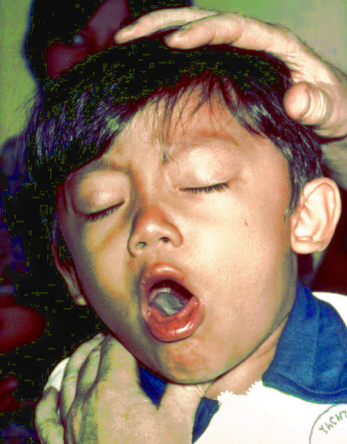
A cough is a sudden, and often repetitively occurring, protective reflex which helps to clear the large breathing passages from fluids, irritants, foreign particles and microbes. The cough reflex consists of three phases: an inhalation, a forced exhalation against a closed glottis, and a violent release of air from the lungs following opening of the glottis, usually accompanied by a distinctive sound.
Cough is the most common reason why patients seek medical attention. Acute cough caused by viral upper respiratory tract infection is usually self-limiting and seldom requires investigation. In contrast, chronic cough is challenging to manage as it often remains unexplained despite thorough investigation.
Causes
- Infective Disorders of Airway
- Common cold
- Sinusitis (post-nasal discharge)
- Tonsillitis, Pharyngitis, Laryngitis
- Laryngotracheobronchitis, Bronchiolitis
- Pneumonia
- Measles, whooping cough
- Inflammatory Disorders of Airway
- Asthma and Loeffler’s syndrome, TPE
- Inhalation of environmental irritants like tobacco, smoke dust.
- Pleural pathology
- Pleural effusion
- Empyema
- Suppurative Lung Disease
- Bronchiectasis
- Cystic fibrosis
- Foreign body retained in bronchi
- Lung abscess
- Anatomic Lesions
- Congenital malformations, Sequestrated lobe
- Bronchomalacia, Tumors
- Tracheal stenosis, H-type TEF
- Vascular ring
- Tracheomalacia
- Irritative
- Post-nasal discharge
- Sinusitis
- GERD
- Irritation of EAM
- Psychogenic
- Habitual cough
- Interstitial Lung Disease
- Compression of Airways
- Lymph nodes
- Retropharyngeal abscess
- Mediastinal mass
- Non-pulmonary causes
- CCF, pericardial effusion, constrictive pericarditis
- Congenital heart diseases.
- Abdominal Causes
- Diaphragmatic hernia, eventration of diaphragm, intra-abdominal masses
- Massive ascites
Life-threatening causes
- Croup
- Laryngeal edema
- FB
- Pertussis
- Bronchiolitis
- Asthma
- Pneumonia
- Toxic inhalation
- CCF
Respiratory sounds
| Sound | Causes | Character |
| Snoring | Oropharyngeal obstruction | Inspiratory, low-pitched irregular |
| Grunting | By partial closure of the glottis | Expiratory, occurs in hyaline membrane disease |
| Rattling | Secretions in trachea/ bronchi | Inspiratory, coarse |
| Stridor | Obstruction larynx/ trachea | An inspiratory sound may be associated with an expiratory component |
| Wheeze | Lower airway obstruction | Continuous musical sound expiratory in nature |
History
- Acute /chronic
- Considered to be chronic if > 2-3 weeks.
- Significant overlap.
- Age of patient
- Infancy- GERD, swallowing dysfunction, CCF associated with CHD.
- Toddlers- RAD, passive smoking, ciliary dyskinesia.
- Adolescents – infections (TB), RAD, psychogenic.
- Past history
- URTI- Post-infectious, irritative, sinusitis.
- H/o contact with TB.
- Associated symptoms
- Fever, nasal discharge suggest infection.
- Fever with chills or night sweats suggests TB.
- Sputum production indicates bronchiectasis or other lower-airway pathology- with the headache or facial edema – sinusitis
- Quality of cough
- Productive cough suggests lower airway infection, CF/bronchiectasis.
- Barking seal-like cough is usually associated with croup.
- Honking or brassy cough is typical in habitual or psychogenic cough.
- Disease of the smaller airways (eg asthma or bronchiolitis)- high-pitched, “ tight” cough.
- Pattern of cough
- Night time cough suggests Reactive Airway Disease.
- With nighttime/early morning cough, consider sinusitis.
- Paroxysmal with whoop – pertussis
- Seasonal cough suggests an allergy.
- Sputum
- Purulent- suppurative lung disease.
- Mucoid – asthma (yellowish sputum in some cases due to eosinophils).
- Hemoptysis – bronchiectasis, MS, CF or FB.
- History of choking (i.e. Retained FB, etc.)
- FTT/ severe undernutrition
- CF, immunodeficiency.
- Chronic cough– TB, bronchiectasis, pertussis, CF, severe chronic asthma or immunodeficiency syndrome.
- Immunization history DPT, measles, BCG
Physical Examination
- General appearance:-
- Evidence of FTT – consider CF, immunodeficiency.
- Pallor – severe anemia
- Cyanosis – hypoxemia/heart disease.
- Subconjunctival hemorrhage – pertussis
- Tachypnea, use of accessory muscle – respiratory distress.
- Grunting, nasal flaring, head nodding, wheezing
- Stridor, in combination with cough, generally indicates obstruction at the level of larynx or trachea.
- JVP – raised in CCF.
- Vital signs:-
- Fever – infective pathology.
- Pulse – tachycardia–CCF, fever, distress.
- RR- to be counted for 1 minute when the child is calm.
- Clubbing:-
- Bronchiectasis, lung abscess, cystic fibrosis.
- ENT Examination:-
- Nose – nasal discharge – blood stained/ serous/ purulent; DNS.
- Polyps – allergy.
- Pharynx – congested/ grey membrane.
- Tonsils – enlargement/ congestion/pus points.
- Ear – discharge (ASOM), retraction of TM.
- Signs of atopic disease:-
- Eczema, transverse nasal crease, rhinitis, mucosal cobblestoning, injected conjunctivae – consider RAD, allergy.
- Sinusitis: – Periorbital edema, sinus tenderness, purulent posterior pharyngeal drainage, halitosis
System Examination
Respiratory system:-
Inspection
- Intercostal or subcostal indrawing.
- Intercostal fullness (pleural effusion, empyema) or crowding of ribs (collapse, fibrosis).
- Decreased movement of either hemithorax.
- Suprasternal recessions – suggestive of narrowing or obstruction of upper airways (laryngeal diphtheria, acute laryngotracheobronchitis, laryngeal/tracheal FB and angioneurotic edema).
- The position of the trachea (trail sign).
Palpation
- Feel for abnormal vibrations – rhonchi, friction rub, crackles, crepitus (subcutaneous emphysema- pertussis).
- Vocal fremitus.
- Expansion of hemithorax.
Percussion
- Area of dullness- pleural effusion (shifting dullness), empyema, consolidation.
- Hyper-resonant – pneumothorax.
- Percuss for upper margin of liver dullness.
Auscultation
- Compare air entry B/L.
- Bronchial breath sounds.
- Added sounds – crackles, rhonchi, pleural friction rub.
- Vocal resonance
- Absent/ decreased – pleural effusion, atelectasis.
- Increased – consolidation, atelectasis with patent bronchus.
Never forget to examine:
- Cardiovascular System
- Abdomen
Investigations
- Complete blood count
- Hb -anemia
- Total and differential count- infections.
- Eosinophilia – TPE, Loeffler’s syndrome.
- Sputum
- AFB
- Eosinophils suggest an asthmatic process or hypersensitivity process of the lung.
- PMN cells suggest infection.
- Lipid laden macrophages- suggest recurrent aspiration.
- Routine or special cultures based on likely pathogens.
- Pulse Oximetry
- For bedside evaluation/monitoring hypoxia.
- Blood C/S
- Important in any infective pathology.
- ABG
- Estimation of partial pressures of O2 and CO2 in blood along with blood PH is used for making the diagnosis of respiratory failure or metabolic acidosis (sepsis/shock)
- X- rays
- CXR – especially in heart diseases, pneumonia not resolving with treatment, pleural effusion.
-Infiltrates may suggest pneumonia, bronchiolitis, pneumonitis, TB, CF, bronchiectasis.
-Volume loss may be seen with foreign body aspiration.
-Hyperinflation suggests RAD or CF.
-Mediastinal nodes may indicate infection (esp. TB, fungus) or malignancy.
- Sinus films – sinusitis.
- Lateral neck X-rays – acute epiglottitis, retropharyngeal abscess.
- Barium swallow –TEF, GERD.
- CT – Scan
- Bronchiectasis (HRCT).
- Lymph nodes, pleural pathologies.
- Pulmonary Function Tests
- To diagnose and follow the course of chronic respiratory illness.
- Immune workup
- Ig levels
- HIV testing
- Bronchoscopy:-
- To remove a foreign body or obtain samples (BAL).
Please click on share to help us grow!

